Institute of Genomic Medicine
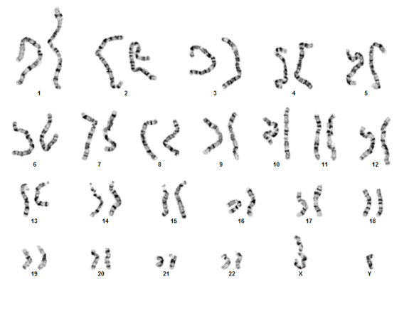
Blood Chromosome Analysis
BACKGROUND:
Cells from peripheral blood are used most often in chromosome analysis, although other cell types can also yield good results. Cells from peripheral blood have several advantages: they are easy to collect, can be cultured rapidly (in three days or less), and only a few drops of blood are necessary for analysis.
INDICATIONS FOR TESTING
Clinical uses of stimulated peripheral blood cultures include:
- Determination of the constitutional karyotype of patients requiring genetic diagnosis or genetic counseling;
- Determination of the constitutional karyotype of families of individuals with chromosome abnormalities to determine carrier status;
- Determination of the constitutional karyotype of leukemia patients who have chromosome abnormalities in their bone marrow;
- Assessment of chromosome damage in individuals exposed to environmental hazards.
SAMPLE REQUIREMENTS
Five milliliters of whole blood collected by venipuncture into a green top sodium heparin tube. In newborns, 1-2 mls of blood may be sufficient.
SPECIMEN HANDLING
Blood may be stored at room temperature overnight, refrigerated up to three days, but never frozen. Blood may also be mailed by overnight courier at ambient temperature. Blood drawn more than five days prior to receipt, or in the wrong tube (such as lithium heparin), is unacceptable.
PROCEDURE
The blood is cultured for 72 hours in culture media with fetal bovine serum, L-glutamine and PHA (phytohemagglutanin) at 5% CO2 and 37C. After culture, blood is exposed to colcemid (to collect mitotic cells), hypotonic solution and fixative, and the cell suspension is dropped on a slide. Slides are G-banded, and 20 cells and two karyotypes are prepared from each routine case.
INTERPRETATION
Peripheral blood analysis will detect numerical chromosomal abnormalities, such as trisomies, monosomies and chromosomal mosaicism; structural abnormalities such as translocations, inversions, derivative chromosomes and additional abnormal chromosomes. Deletions and rearrangements on the molecular level cannot be seen. The origin and structure of derivative and structurally abnormal chromosomes can be deduced by following G-banding analysis by fluorescent in situ hybridization (FISH).
Microdeletion syndromes can be detected by FISH analysis (see FISH section).
RESULTS
Results are presented according to the international standards for chromosomal nomenclature (ISCN), and a full explanation of the karyotype and clinical implications is provided. Any abnormal or variant results are followed by genetic counseling.